Back Of Skull Anatomy - Posterior And Lateral Views Of The Skull Anatomy Kenhub - Skull bones aren't fused together at birth.. Excluding ear ossicles, it is made of 22 bones. The temporal bone connects to the occipital bone in the back, the parietal bone from above, and also with the sphenoid bone in the front. Anatomical structures of the skull include: The skull also includes cartilage (put your finger on the tip of your nose and wiggle it) and ligaments (open and close your mouth if you want to use them). Learn about the anatomy of the skull bones and sutures as seen on ct images of the brain.
Anatomical structures of the skull include: William is a final year medical student in australia who has taught anatomy to tertiary science and. The skull also includes cartilage (put your finger on the tip of your nose and wiggle it) and ligaments (open and close your mouth if you want to use them). It offers protection to the brain, eye balls, inner ears, and nasal passages. Foramina inside the body of humans and other animals.
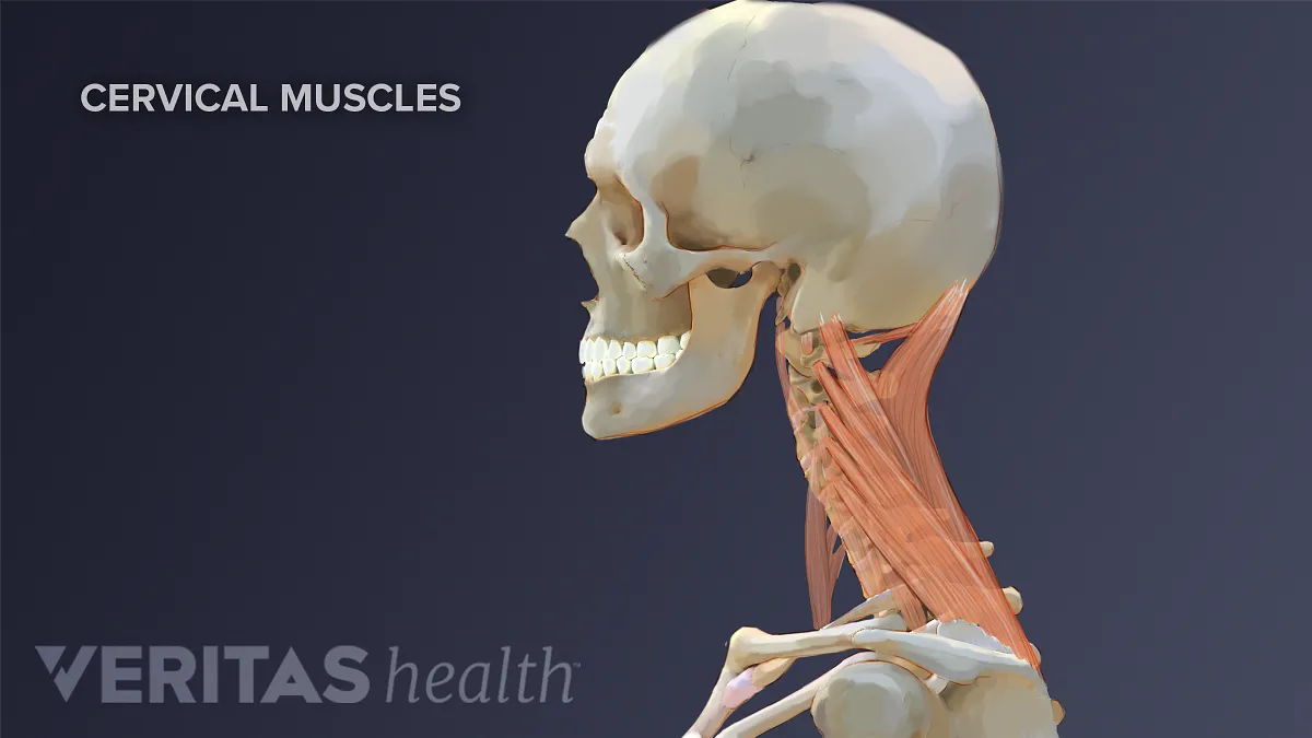
Neck Muscles And Other Soft Tissues from embed.widencdn.net The major sutures are the coronal suture, sagittal suture, lambdoid suture and squamosal sutures. A cartilaginous mould begins to grow this is why raising your eyebrows can create the appearance that the back of the head is moving. The skull includes the upper jaw and the cranium. A thorough description is beyond the. The cranium and mandible was exported from ct data. The occipital bone forms the back of the skull and the base of the cranium. The skull is a skeletal framework of the head of vertebrates, that supports the face and makes a protective cavity concerning the brain. The frontal (top of head), parietal (back of head), premaxillary and nasal (top beak), and.
Overview, anterior skull base, middle skull base march 18, 2017.
The bbc is not responsible for the content of external websites. William is a final year medical student in australia who has taught anatomy to tertiary science and. Overview, anterior skull base, middle skull base march 18, 2017. It offers protection to the brain, eye balls, inner ears, and nasal passages. Cranium) is the skeleton of the head composed of 22 separate bones joined together primarily by sutures. The skull bones can be classified into two groups: In order to be light, the skull is made up by flat and irregular bones, and has hollow spaces called the sinuses. Learn more about the anatomy and function of the skull in humans and other vertebrates. Learn vocabulary, terms and more with flashcards, games and other study tools. The base of the skull (or skull base) forms the floor of the cranial cavity and separates the brain from the structures of the neck and face. The occipital bone forms the back of the skull and the base of the cranium. Learn about the anatomy of the skull bones and sutures as seen on ct images of the brain. Anatomy and physiology7.2 the skull.
From an anatomical perspective, the skull is divided into two parts: The base of the skull (or skull base) forms the floor of the cranial cavity and separates the brain from the structures of the neck and face. Anatomical structures of the skull include: A cartilaginous mould begins to grow this is why raising your eyebrows can create the appearance that the back of the head is moving. Learn about the anatomy of the skull bones and sutures as seen on ct images of the brain.
Bones Of The Head Atlas Of Anatomy from doctorlib.info Skull reshaping is done on any of the structures that lie above the face. Excluding ear ossicles, it is made of 22 bones. Anatomy of the skull and bones of cranium on medical illustrations. Learn about the anatomy of the skull bones and sutures as seen on ct images of the brain. The frontal, parietal, temporal and occipital bones are joined at the cranial sutures. It is comprised of many bones, formed by intramembranous ossification, which are joined together by sutures (fibrous joints). Human skull from the front. The skull has a single occipital condyle.7 the skull consists of five major bones:
It is comprised of many bones, formed by intramembranous ossification, which are joined together by sutures (fibrous joints).
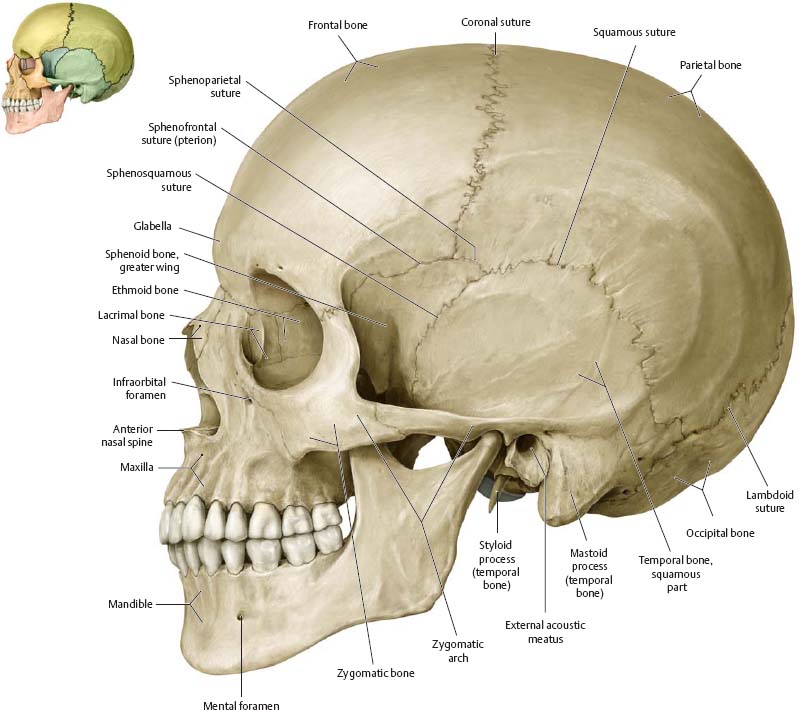
The cranium and mandible was exported from ct data. Learn more about the anatomy and function of the skull in humans and other vertebrates. The cranium and the mandible. Human skull from the front. Looking at it from the inside it can be subdivided into. In order to be light, the skull is made up by flat and irregular bones, and has hollow spaces called the sinuses. These joints fuse together in adulthood. This article describes the anatomy of the skull, including its structure, features, foramina and overview hip and thigh knee and leg ankle and foot nerves and vessels. Some bones give shape to the face, others protect the brain. The axial & appendicular skeleton. They don't move and united into a single unit. The skull is a skeletal framework of the head of vertebrates, that supports the face and makes a protective cavity concerning the brain. Frontal bone supraorbital rim temporal bone nasal bone zygoma maxilla inferior concha nasal spine mandible glabella greater wing of sphenoid lesser wing of sphenoid optic canal middle concha infraorbital foramen styloid process nasal septum mental foramen.
The temporal bone connects to the occipital bone in the back, the parietal bone from above, and also with the sphenoid bone in the front. This is a model of the human (homo sapiens) skull. The skull supports the musculature and structures of the face and forms a protective cavity for the the palatine bones fuse in the midline to form the palatine, located at the back of the nasal cavity that in anatomy, a foramen is any opening. The bone is pierced by a large oval hole(the foramen magnum) through which runs the spinal cord. A thorough description is beyond the.
Cranial Bones Function And Anatomy Diagram Conditions Health Tips from post.healthline.com The skull is a skeletal framework of the head of vertebrates, that supports the face and makes a protective cavity concerning the brain. The skull or known as the cranium in the medical world is a bone structure of the head. They don't move and united into a single unit. Skull, skeletal framework of the head of vertebrates, composed of bones or cartilage, which form a unit that protects the brain and some sense organs. Anatomy and physiology7.2 the skull. The axial & appendicular skeleton. From an anatomical perspective, the skull is divided into two parts: Learn about the anatomy of the skull bones and sutures as seen on ct images of the brain.
Skull bones aren't fused together at birth.
Anatomy and physiology7.2 the skull. Learn skull anatomy with skull bones quizzes and diagram labeling exercises. Anatomical structures of the skull include: The skull base is the inferior portion of the neurocranium. The occipital bone forms the back of the skull and the base of the cranium. Skull bones aren't fused together at birth. Looking at it from the inside it can be subdivided into. Learn vocabulary, terms and more with flashcards, games and other study tools. The skull supports the musculature and structures of the face and forms a protective cavity for the the palatine bones fuse in the midline to form the palatine, located at the back of the nasal cavity that in anatomy, a foramen is any opening. This is a model of the human (homo sapiens) skull. The skull has a single occipital condyle.7 the skull consists of five major bones: It is comprised of many bones, formed by intramembranous ossification, which are joined together by sutures (fibrous joints). Some bones give shape to the face, others protect the brain.


0 Komentar